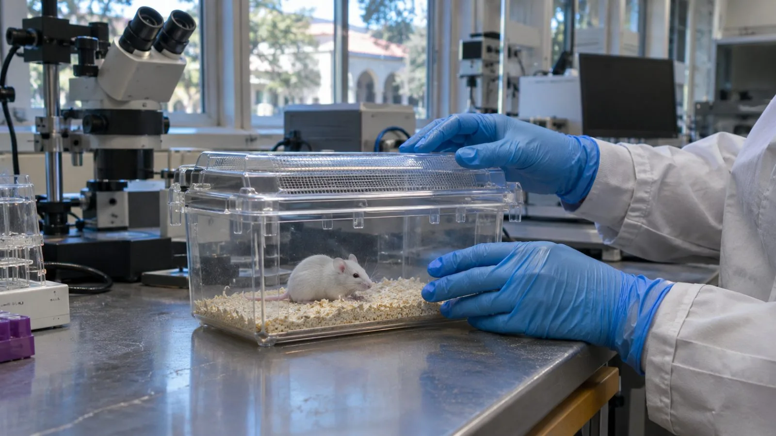
In seven genetically engineered mice, transplanted human-derived tissue accounted for 91.9% of combined cortical volume after three months, testing a living model for research into brain development and injury.
Published Sept. 16 in Nature, the study involved four cortical organoids per mouse. The organoids were 30 to 60 days old when transplanted; the recipient pups were 5 to 17 days old, with a median age of 10 days. The animals had been engineered to lack most of the neocortex and hippocampus.
MRI detected grafts in 86.2% of 29 mice at the two-month scan. In a separate group of 14 animals, graft volume rose 4.7-fold between months two and three.
The grafts contained human cortical cell types and showed organized electrical activity, the study reports. Human-derived projections reached parts of the mice’s nervous systems, including the spinal cord. The paper leaves open whether electrical activity from the graft also reached host circuits.
In a Y-maze, both the control group and grafted XCX animals performed better than chance; ungrafted apallial, or cortex-depleted, mice did not. The authors say the results do not establish whether graft-derived neurons were necessary or sufficient for any particular behavior.
The team also examined low-oxygen injury. Researchers gradually reduced oxygen for an hour, then exposed the mice to 5% oxygen for five hours. Two days later, grafted mice showed changes in gait measures. The study presents this as a model for examining injury to developing cortical tissue; it did not test a treatment.
Organoids grown in dishes lack the blood supply, sensory signals and wider neural connections found in a living nervous system, the paper says. Sergiu P. Pașca, the study’s senior author and a Stanford professor of psychiatry and behavioral sciences, said the approach could help researchers see how disease-related changes affect human neural networks in a living animal.
The mice underwent genetic cortical depletion, surgery and behavioral testing; some were also exposed to low oxygen. The paper reports animal-welfare monitoring, review by Stanford stem-cell and animal-care committees, bioethicist consultation and guidance from an external independent ethics committee. Cell donors consented to transplantation research. The authors call for ethics input before studies extend graft maturation or involve nonhuman primates.