Researchers in Japan used high-speed imaging to track how a chromosome protein complex changes shape and holds two DNA segments together, clarifying a possible mechanism for organizing genetic material.
In a laboratory tethering test, about 25% of free DNA added to a sample was recovered when adenosine triphosphate, or ATP, was present. Recovery fell below 3% when researchers substituted adenosine diphosphate, or ADP, or left ATP out. The test provided a separate measure of the complex’s ability to bring DNA segments together. Researchers ran it for 30 minutes at 30 degrees Celsius and reported three independent experiments.
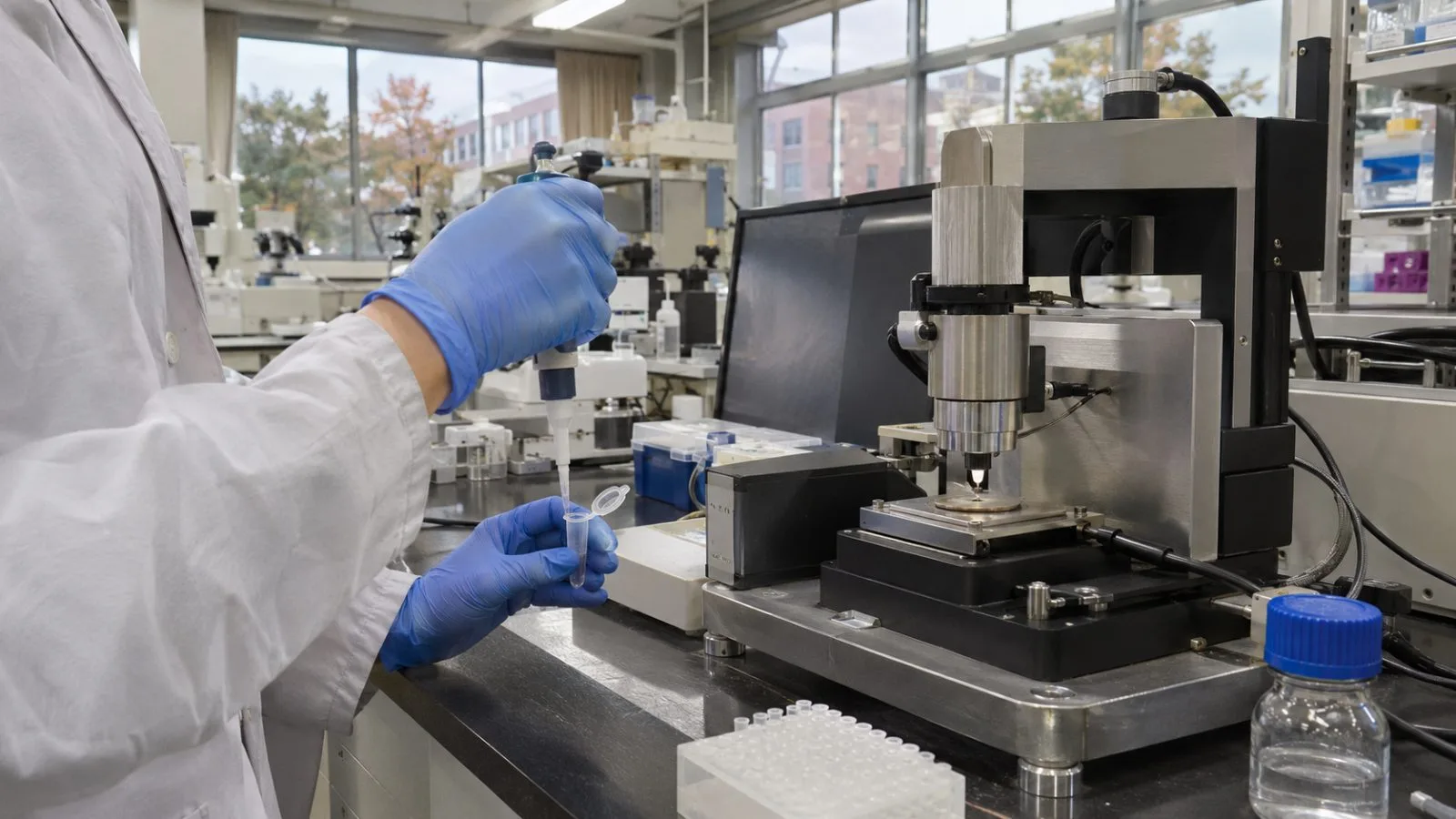
Kanazawa University announced the findings Nov. 4, following their Sept. 1 publication in Nature Communications. Its researchers and colleagues at the National Institute of Genetics studied Smc5/6 purified from budding yeast. They used high-speed atomic force microscopy to record the complex as it interacted with DNA.
When ATP bound to Smc5/6, its two arms spread from a closely aligned position into a more open shape. DNA initially contacted the ends where the complex processes ATP. After the ATP was broken down, the DNA occupied space inside the ring, nearer the hinge connecting its arms. The observations link changes in the complex’s shape to different positions for the DNA.
The images also showed Smc5/6 holding two stretches of DNA at once and maintaining twisted DNA in a more compact arrangement. For twisted circular DNA, the mean radius of gyration was about 55 nanometers, compared with about 80 nanometers for DNA without Smc5/6.
Smc5/6 belongs to a family of ring-shaped complexes involved in organizing chromosomes. An earlier study in Nature reported that Smc5/6 can draw DNA into loops. The new experiments used purified material outside living cells, so they do not establish the full sequence of events in a cell or demonstrate a medical application.